
Does Electroencephalography Enable a Future Where Mind Reading is Possible?
EEG technology has been available for almost 100 years, yet we we may have not yet unlocked its full potential.
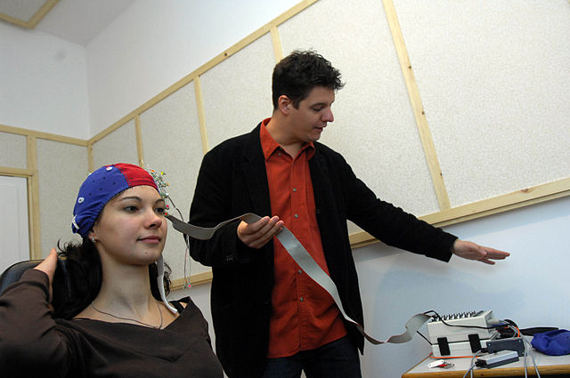
Csaba Segesvári camera-man at Délmagyarország lapcom Kft., CC BY-SA 3.0
Electroencephalography allows us to detect brain abnormalities in people, as it is low-cost, non-invasive, and safe. Its usage may become widespread for early detection of diseases and disorders.
Electroencephalography (EEG) may be the future, allowing people to tap into their own minds with low-risk, non-invasive, and low-cost EEG technology. It would allow us to better understand brainwaves, make our lives easier and prevent growth in neurological abnormalities.
Electroencephalography technology uses electrodes to measure the signals of our brain’s waves and activities. It is most commonly used to detect brain abnormalities and can diagnose various diseases such as Alzheimer’s disease, CNS disorders, and narcolepsy. There are various parts of this scan, as EEG examines our brain’s alpha, beta, theta, delta, and gamma waves, of which each have different frequencies.
Though it cannot directly read a person’s mind, it is one of the most effective ways to be at least close to being able to do so while keeping costs low. The benefit of using EEG technology is that it can allow us to detect issues within the brain early, especially when used in a widespread level.
Recent research has shown us how life flashes before our eyes with our memories from throughout the lifetime when we take our last breath. This active impulse from our brain has allowed researchers to uncover what this experience is like, thanks to EEG technology being used to measure the brain activity right before an 87-year old Canadian man’s heart finally stopped beating leading to his death. This was the first time we have ever had a recorded electroencephalography scan during a person’s death, as he had died during a surgical procedure for his epilepsy and had an unexpected heart attack while the brain scanner was still running.
“This is why it’s so rare, because you can’t plan this. No healthy human is going to go and have an EEG before they die, and in no sick patient are we going to know when they’re going to die to record these signals,” study author Ajmal Zemmar, a neurosurgeon at the University of Louisville in Kentucky, said to Insider’s Anna Medaris Miller.
This scan showed that the brain is likely to replay the nicest memories in a person’s life when we die. It also showed that even after the heart stops beating, there was continuous brain activity for a moment that showed memory recall taking place. Prior to this, there were no confirmations of life flashing before our eyes with brain scanning in place. However, some limitations to this include that there may have been altered gamma waves due to his epilepsy.
This work has gone even further with research at the University of Toronto by Dan Nemrodov, where he led the work in digitally reconstructing images inside the mind with the data captured from electroencephalography.
“When we see something, our brain creates a mental percept, which is essentially a mental impression of that thing. We were able to capture this percept using EEG to get a direct illustration of what’s happening in the brain during this process,” said Dan Nemrodov to the University of Toronto.
Test volunteers were shown photos of faces while connected to EEG equipment for the study. Their brain activity was recorded, and a technology based on machine learning algorithms was utilized to digitally reproduce the image in the subject’s head.
Image reconstruction has been made possible in the past through fMRI, especially with facial images. Despite the advancements, fMRI falls short in long-term practicality. However, fMRI is not portable and is very expensive. While it provides a better spatial resolution of the brain, it only provides a snapshot, unlike the EEG which provides a real-time analysis down to the second. This experiment was the first time this was done with the EEG rather than fMRI.
Researchers had doubted the ability of EEG technology to be able to do such a thing; however, this experiment had shown the great practical potential of this technology being used at a widespread level with various purposes in uncovering the various parts of the brain.
“The fact we can reconstruct what someone experiences visually based on their brain activity opens up a lot of possibilities. It unveils the subjective content of our mind, and it provides a way to access, explore and share the content of our perception, memory, and imagination,” said Dr. Adrian Nestor from the University of Toronto, who is the Director of the Visual Recognition Laboratory.
“When we see something, our brain creates a mental percept, which is essentially a mental impression of that thing. We were able to capture this percept using EEG to get a direct illustration of what’s happening in the brain during this process,” said Dan Nemrodov to the University of Toronto.
Ryan Ahmed is a Managing Editor for 'The Science Survey.' He wants to uplift communities and provide unique insight on current events around the globe...